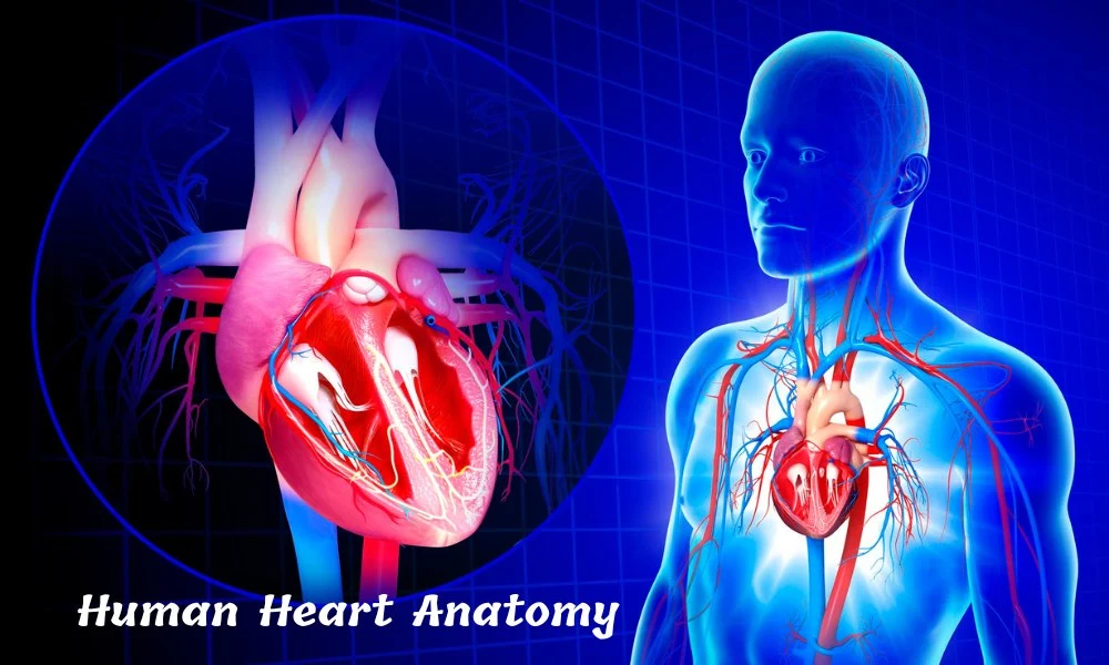
The human heart is an extraordinary organ, integral to our survival by continuously pumping blood throughout the body. Grasping the intricacies of heart anatomy is essential for understanding its vital functions and maintaining optimal heart health. This article provides an in-depth overview of the human heart anatomy, including heart structure, layers of the heart, heart chambers and valves, and Cardiac Cycle. Learn about common heart diseases and tips for maintaining heart health in this comprehensive guide.
Introduction to Human Heart Anatomy

The human heart is the body’s central pump, crucial for sustaining life by delivering oxygen-rich blood to various tissues and organs. This guide provides an in-depth look at the heart’s anatomy, highlighting its key structures and functions.
Location and Structure of the Heart
The heart is situated in the thoracic cavity, nestled between the lungs and slightly offset to the left. It is protected by the ribcage and lies above the diaphragm.
Layers of the Heart

The heart’s wall is composed of 4 main layers:
- Pericardium: A double-walled sac that encloses and protects the heart, reducing friction during heartbeats and its movements.
- Epicardium: The thin outermost layer of the heart wall.
- Myocardium: Thick, muscular middle layer responsible for the heart’s pumping action.
- Endocardium: The innermost layer that lines the heart chambers and valves, ensuring smooth blood flow.
(Also Read: Exploring the Different Types of Skin Layers: A Complete Overview…)
Heart Chambers and Valves

The human heart comprises four chambers: two atria and two ventricles.
Atria
- Right Atrium: Receives deoxygenated blood from the body via the superior and inferior vena cava.
- Left Atrium: Receives oxygenated blood from the lungs via the pulmonary veins.
Ventricles
- Right Ventricle: Pumps deoxygenated blood to the lungs through the pulmonary artery.
- Left Ventricle: Pumps oxygenated blood to the body through the aorta.
Valves
The heart contains four main valves that ensure one-way blood flow:
- Tricuspid Valve: Located between the right atrium and right ventricle.
- Pulmonary Valve: Situated between the right ventricle and pulmonary artery.
- Mitral Valve: Positioned between the left atrium and left ventricle.
- Aortic Valve: Found between the left ventricle and aorta.
Blood Circulation Through the Heart

Blood circulation through the heart follows a specific pathway to ensure efficient oxygenation and nutrient delivery.
- Deoxygenated Blood Flow:
- Blood enters the right atrium from the body.
- Passes through the tricuspid valve into the right ventricle.
- Is pumped through the pulmonary valve into the pulmonary artery and sent to the lungs for oxygenation.
- Oxygenated Blood Flow:
- Blood returns from the lungs to the left atrium.
- Moves through the mitral valve into the left ventricle.
- Is pumped through the aortic valve into the aorta and distributed throughout the body.
(Also Read: Top 10 Hormone Balancing Foods for Optimal Health….)
Coronary Arteries and Blood Supply
The heart itself requires a dedicated blood supply, provided by the coronary arteries. These arteries branch from the aorta and encircle the heart, delivering oxygen-rich blood to the myocardium.
Major Coronary Arteries
- Left Coronary Artery (LCA): Divides into the left anterior descending artery (LAD) and the circumflex artery (LCX), supplying the left side of the heart.
- Right Coronary Artery (RCA): Supplies the right side of the heart and portions of the left ventricle.
The Electrical System of the Heart
The heart’s electrical system controls its rhythm and ensures coordinated contractions. Key components include:
- Sinoatrial (SA) Node: The heart’s natural pacemaker located in the right atrium, initiating electrical impulses.
- Atrioventricular (AV) Node: Receives impulses from the SA node and transmits them to the ventricles.
- Bundle of His: Conducts impulses from the AV node to the ventricles.
- Purkinje Fibers: Distribute electrical impulses throughout the ventricles, triggering contractions.
Heart Function and Cardiac Cycle
The cardiac cycle consists of two main phases: systole and diastole.
- Systole: The phase of contraction, where the ventricles pump blood into the pulmonary artery and aorta.
- Diastole: The phase of relaxation, where the heart chambers fill with blood.
This cycle ensures continuous blood flow, maintaining the body’s vital functions.
(Also Read: Parts Of The Brain And Their Functions…)
Common Heart Diseases and Conditions
Understanding heart anatomy is crucial for recognizing common heart diseases and conditions, such as:
- Coronary Artery Disease (CAD): Blockage of coronary arteries, leading to reduced blood flow to the heart muscle.
- Myocardial Infarction (Heart Attack): This occurs when a coronary artery is completely blocked, causing heart tissue damage.
- Heart Failure: The heart’s inability to pump blood effectively, resulting in fluid buildup and fatigue.
- Arrhythmias: Irregular heartbeats caused by issues in the heart’s electrical system.
- Valvular Heart Disease: Malfunctioning heart valves that disrupt blood flow.
Maintaining a Healthy Heart
Keeping your heart healthy is essential for a long and active life. Here are some tips for heart health:
- Healthy Diet: Consume a balanced diet rich in fruits, vegetables, whole grains, and lean proteins.
- Regular Exercise: Engage in at least 150 minutes of moderate aerobic activity or 75 minutes of vigorous activity per week.
- Avoid Smoking: Smoking is a major risk factor for heart disease.
- Manage Stress: Practice stress-reducing techniques such as meditation, yoga, or deep breathing.
- Regular Check-ups: Visit your healthcare provider for regular heart health screenings and blood pressure monitoring.
Conclusion
The human heart is a complex and vital organ, central to sustaining life. Understanding heart anatomy, from chambers and valves to its electrical system, is essential for appreciating how it functions and how to maintain heart health.